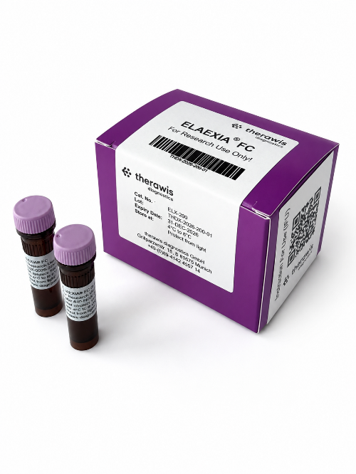
The ELAEXIA® FC detects exosomes in biofluids such as cell culture supernatant or human plasma. The detection is performed by means of flow cytometry applying fluorescently labelled ELAEXIA® FC which binds specifically with high affinity to the lipid membrane of exosomes
- Catalog no.: ELX-200-01
- List price: €480.00 net · excl. shipping
- Product name: ELAEXIA® FC
- Size: 40 detections
- Quantity: 200µl
For research use only!
Order here

Instruction for Use
Safety Data Sheet
Background
Extracellular vesicles (EVs) are generated almost by any cell and different subcellular compartments and, therefore, EVs display a significant heterogeneity and diversity. Flow cytometry analysis using fluorescently labelled antibodies against the exosome markers CD9, CD81, or CD63 will detect subpopulations of exosomes because the extracellular versicles are loaded with different protein cargo depending on the state of the cells they originate from. Our novel, highly selective method is based on the exosome biogenesis. Exosome biogenesis occurs in cells and at the molecular level inside the Multi-Vesicular Body (MVB) compartment. The donor membrane for the budding vesicle is the MVB membrane (Subra et al., 2007). Lipids such as phosphatidylserine and phosphatidylethanolamine, usually in the inner leaflet of the membrane of cells, form through this inward budding of MVBs the outer leaflet of the exosome membrane. ELAEXIA®, Elective Labelling of Exosomes for Identification and Analysis, binds specifically to phosphatidylethanolamine in the exosome lipid bilayer membrane.
Key features of ELAEXIA® FC are:
- Precise and specific binding to exosome membrane bilayer potentially identifying all exosomes
- This novel technology is independent of highly variable protein markers which identify only exosome subpopulations
- Direct exosome labelling in cell culture supernatant and plasma for multiparametric flow cytometry analysis
- Highly reproducible results (intra- and inter-assay CV %)
- Compatible with antibody co-staining and multiparametric analysis
- Labelling of exosomes for immunofluorescence microscopy, Nanoparticle Tracking Analysis (NTA) and nano imager analysis
- Fast and Easy-to-use labelling protocol
Data and images
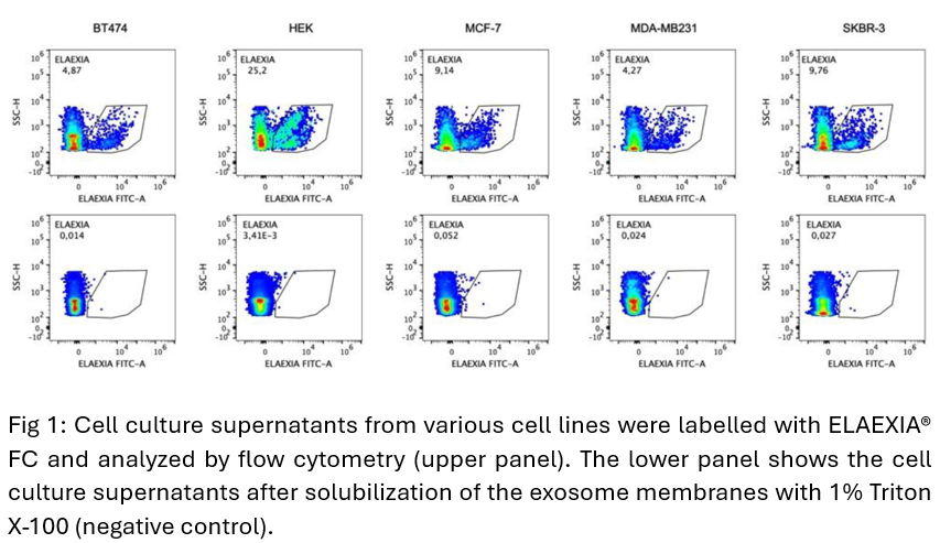
ELAEXIA® FC stains by flow cytometry analysis extracellular vesicles from various cell lines.
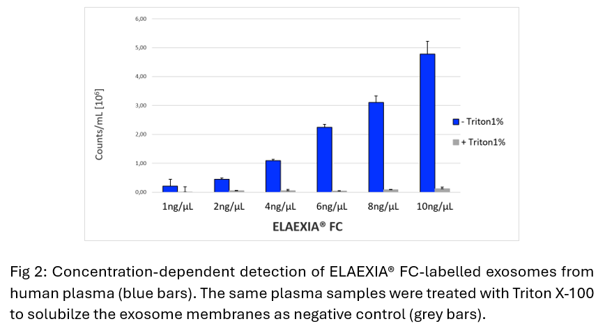
ELAEXIA® FC detects exosomes in human plasma by flow cytometry.